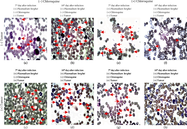
Figure 3.

Photomicrography obtained from blood smear on days 7 and 10 after Plasmodium berghei ANKA infection. In (a)–(d), mice were not treated with chloroquine. In (e)–(h), the mice were treated with chloroquine. In (c) and (d) and (g) and (h), mice were inoculated with the Ehrlich tumor. Red arrows indicate ring-form trophozoites. Photomicrography was obtained for an optic microscope (NIKON Ni-E) at 100x magnification.
