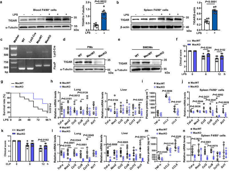
Fig. 1. Ablation of Tigar ameliorates inflammation in murine sepsis.
a, b Male C57BL/6 J mice were intraperitoneally injected with LPS (10 mg kg−1) or phosphate-buffered saline (PBS). The mice were euthanized 12 h later. Western blot analysis of TIGAR expression in F4/80+ cells isolated from both blood (n = 6) (a) and spleen (n = 7) (b). c Representative DNA gel image of WT, Lyz2-Cre, MacWT (Tigarflox/flox), and MacKO (Tigarflox/floxLyz2-CreKI/KI) mice by PCR amplification. The experiment was repeated three times independently with similar results. d, e Western blot of TIGAR in murine peritoneal macrophages (PMs) (d) and BMDMs (e). Blot assay was repeated three times independently with similar results. f Mice were intraperitoneally injected with LPS (10 mg kg−1) and euthanized 12 h later. Clinical score was determined at the indicated time (n = 7). g Murine survival rate was determined during 96 h of LPS challenge (n = 12). h mRNA levels of pro-inflammatory genes in the murine lung (left) and liver (right) after 12 h of LPS challenge (n = 7). i Plasma levels of TNF-α, C–C motif ligand-2 (CCL2) and C–C motif ligand-8 (CCL8) in septic mice (n = 7). j mRNA levels of pro-inflammatory genes in the spleen F4/80+ cells from MacWT (n = 6) and MacKO (n = 7) mice. k Male mice were induced CLP sepsis and euthanized 12 h later. Clinical score was determined at the indicated time (n = 8). l mRNA levels of pro-inflammatory genes in the murine lung (left) and liver (right) after 12 h of CLP surgery (n = 7). m Plasma levels of TNF-α, CCL2 and CCL8 in CLP septic mice (n = 8). n mRNA levels of pro-inflammatory genes in the spleen F4/80+ cells from MacWT and MacKO (n = 7) mice. Data are expressed as mean ± SEM. a Two-tailed Mann–Whitney U test. b, f, k Two-tailed Student t-test. g Log-rank (Mantel–Cox) test. h–j, l–n Two-tailed Student t-test, Two-tailed Mann–Whitney U test. Source data are provided as a Source Data file.

