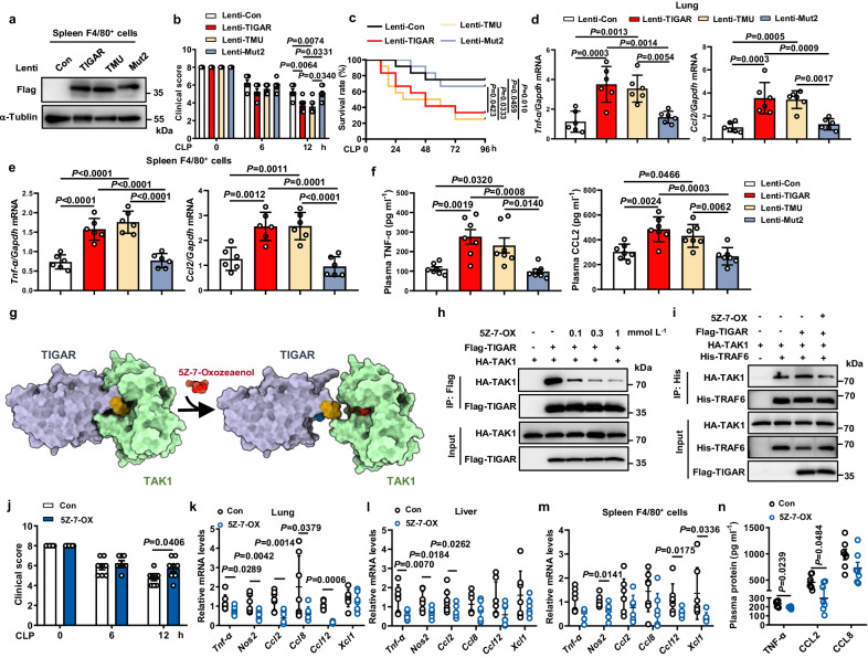
Fig. 7. Inhibiting TIGAR-TAK1 interaction abolishes murine sepsis.
Tigar KO mice were infected with myeloid-specific CD11b promoter-driven lentivirus (pCDH-CD11b-T2A-copGFP) encoding Flag-TIGAR, Flag-TMU, or Flag-Mut2, respectively. After 4 days the CLP sepsis model was generated. a Western blot of Flag-TIGAR, Flag-TMU and Flag-Mut2 expression in spleen F4/80+ cells. b Clinical score of mice was analyzed (n = 7). c Murine survival rate was determined during 96 h of CLP challenge (n = 12). d, e mRNA levels of pro-inflammatory genes in lung (d) (n = 6), and spleen F4/80+ cells (e) (n = 6). f Plasma concentrations of TNF-α, and CCL2 in mice (n = 7). g Schematic diagram of the mechanism underlying 5Z-7-OX competing with TIGAR for binding to TAK1. Yellow structure: residues 152–157 of TIGAR; Blue structure: residues 158–161 of TIGAR. h HEK293 cells were transfected by HA-TAK1 and Flag-TIGAR plasmids. Co-IP and western blot of TIGAR-TAK1 complex formation in HEK293 cells incubated with 0.1, 0.3, or 1 mM 5Z-7-OX for 12 h. i HEK293 cells were transfected by HA-TAK1, His-TRAF6 and Flag-TIGAR plasmids and treated with or without 0.1 mM 5Z-7-OX. Co-IP and western blot of TAK1-TRAF6 complex formation in HEK293 cells. Male C57BL/6J mice were intraperitoneally injected with 5Z-7-OX or DMSO (Con). After 1 h, mice were induced with CLP sepsis and euthanized 12 h later. j Clinical score of mice was analyzed (n = 8). k–m mRNA levels of pro-inflammatory genes in lung (k) (n = 7), liver (l) (n = 7), and spleen F4/80+ cells (m) (n = 7). n Plasma concentrations of TNF-α, CCL2 and CCL8 in Con and 5Z-7-OX (n = 7) treated mice. Data are expressed as mean ± SEM. b, d–f One-way ANOVA followed by the Bonferroni test. c Log-rank (Mantel–Cox) test. j Two-tailed Student t-test. k–n Two-tailed Student t-test, Two-tailed t-test with Welch correction, or two-tailed Mann–Whitney U test. Source data are provided as a Source Data file.

