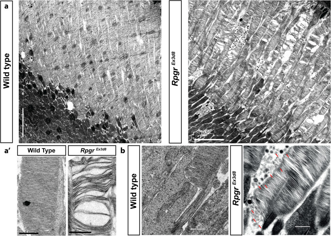
Fig. 2. Mutations in Rpgr lead to early structural compromise of photoreceptor outer segments (OS).
a Transmission electron micrograph of 6 week old wild-type photoreceptors show OS composed of compacted discs extending to the underlying retinal pigment epithelium (RPE) (left panel). Disc compaction is compromised in age-matched mutant photoreceptors, with discs appearing split and spaced out (right panel). a’ Higher magnification examples. b Examination of basal discs at OS base shows compacted discs but an accumulation of shed vesicles (red arrows) in RpgrEx3d8 mice. (Scale bars; a = 5 μm; b = 0.5 μm). (Similar results seen in > 10 experiments repeated independently).

