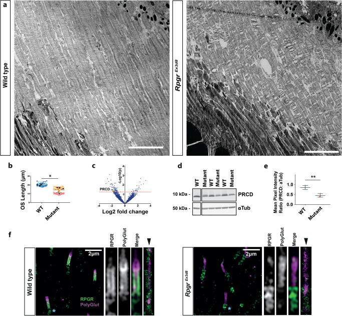
Fig. 3. Loss of Rpgr leads to shortened photoreceptor outer segments (OS).
a Transmission electron micrograph of 6 week wild type and RpgrEx3d8 photoreceptors show OS are shorter in RpgrEx3d8 mice. b Quantification of OS length. Different colours represent measurements from individual mice. Mean OS length measurement of each experimental animal denoted by black circles (WT) and black squares (mutant); N = 3 animals per genotype; *, p = 0.0405; two-tailed, unpaired t-test; error bars denote SEM. c Mass spectrometry comparing protein composition of wild type versus RpgrEx3d8 3 month old retinas shows reduced expression of the outer segment protein PRCD in mutant mice. Red line denotes cut-off p value for significance (two-tailed, unpaired t-test, not corrected for multiple hypothesis testing). d Immunoblotting of whole retina lysates confirms reduced PRCD in mutant mice, in keeping with reduced outer segment lengths. e Quantification of intensity of PRCD relative to loading control α-tubulin; N = 3 animals per group; *p = 0.0062; two-tailed, unpaired t-test). Data presented as mean values +/− SEM. f Left panel: Localisation of RPGR’s retinal-specific isoform throughout the length of the photoreceptor connecting cilium, as evidenced by SIM imaging, showing co-localisation with polyglutamylated tubulin in wild type photoreceptors, which extends the length of the connecting cilium and into the OS63. (Similar results seen in 4 independent experiments using separate animals). RPGR localizes to the ciliary membrane by STORM imaging (rightmost panel, arrowhead; similar results seen in 3 independent experiments). Right panel: RpgrEx3d8 photoreceptors show loss of RPGR staining at the connecting cilium. (NB. green staining below polyglutamylation labelling (see blue *) represents non-specific centrosomal staining; a common occurrence with rabbit polyclonal antibodies) (Scale bars; a = 5 μm; f = 2 μm). Source data provided as a Source Data file.

