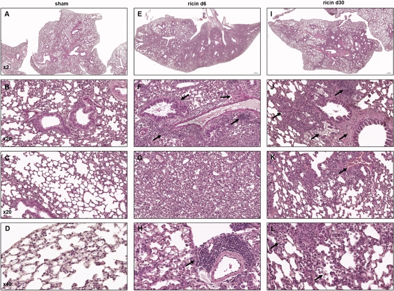
Figure 3.
Histopathological lesions in mice following sublethal ricin intoxication. Paraffin-embedded lung tissues from mice 6 or 30 days following intranasal ricin intoxication (1.7 µg/kg body weight). Lung histopathology from control mice that were intranasally exposed to PBS exhibited normal lung architecture (A–D). Lung tissues from mice 6 days following ricin exposure (E–H). Lung tissues from mice 30 days following ricin exposure (I–L). Black arrows indicate the presented lesions. Panels are representatives of 5 mice in each group, scale bar: 500 µm (A,E,I); 50 µm (B,C,F,G,J,K); 20 µm (D,H,L).

