Figure 5.
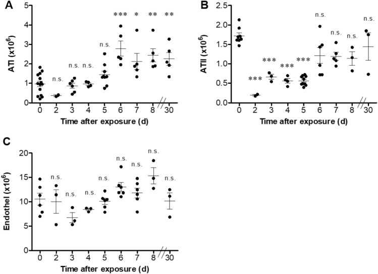
Parenchymal cell alterations following sublethal ricin intoxication. Lungs were isolated from intranasally ricin-intoxicated (1.7 µg/kg body weight) mice at indicated time-points. Lung cell suspensions were stained for (A) ATI, (B) ATII, (C) endothelial cells and analyzed by flow cytometry. Each dot represents single mouse. (n = 3–13). Data are means ± SEM. *P < 0.05, **P < 0.01, ***P < 0.001, n.s., not significant in comparison to non-intoxicated mice.

