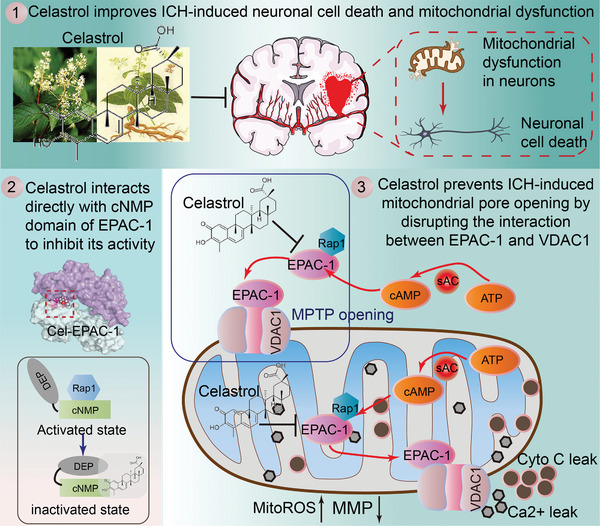
Figure 10.

Graphic illustration of neuroprotective effects and mechanisms of celastrol. Following ICH, EPAC‐1 is activated within neurons and translocated to the outer membrane of mitochondria. Here, it can form a complex with VDAC1, promoting MPTP opening and subsequent collapse of mitochondrial membrane potential. This results in Ca2+ release, which induces neuronal apoptosis via cytochrome C (Cyto C). As a natural compound, celastrol can directly localize in mitochondria and interact with EPAC‐1 to modulate its activation, thereby impeding the binding of EPAC‐1 to VADC1. This ameliorates mitochondrial impairment in neuronal cells and exerts neuroprotective effects after ICH.
