Figure 2.
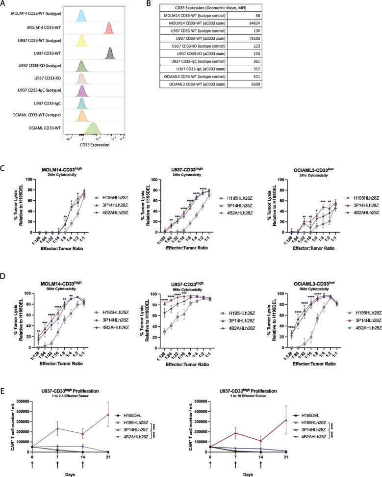
Membrane-proximal targeting chimeric antigen receptor (CAR) T cells demonstrate enhanced functionality and proliferative capacity in vitro. (A) Flow cytometry histograms of CD33 expression on acute myeloid leukemia (AML) cells detected with isotype control or fluorescently labeled CD33-specific antibody. (B) Quantitative geometric mean fluorescence intensity (MFI) of CD33 expression on AML cells detected with either isotype control or fluorescently labeled CD33-specific antibody. (C) 24-hour D-luciferin assay demonstrating lysis of CD33-expressing tumor cells (n=4 biological replicates; ****p<0.0001; ***p<0.001; **p<0.01; *p<0.05 by two-way analysis of variance (ANOVA)). Data are a mean±SEM of four biological replicates. (D) 96-hour D-luciferin assay demonstrating lysis of CD33 expressing tumor cells (n=4 biological replicates; ****p<0.0001; ***p<0.001; **p<0.01; *p<0.05 by two-way ANOVA). Data are a mean±SEM of four biological replicates. (E) Quantification of flow cytometric analysis demonstrating enhanced proliferation by membrane-proximal CD33 targeting CAR T cell in the presence of U937-CD33high tumor cells at E:T ratios of 1:2.5 (left) or 1:10 (right) (n=3 biological replicates; ****p<0.0001 by two-way ANOVA at day 21). Arrows indicate when additional target cells were added. Data are a mean±SEM of three biological replicates.

