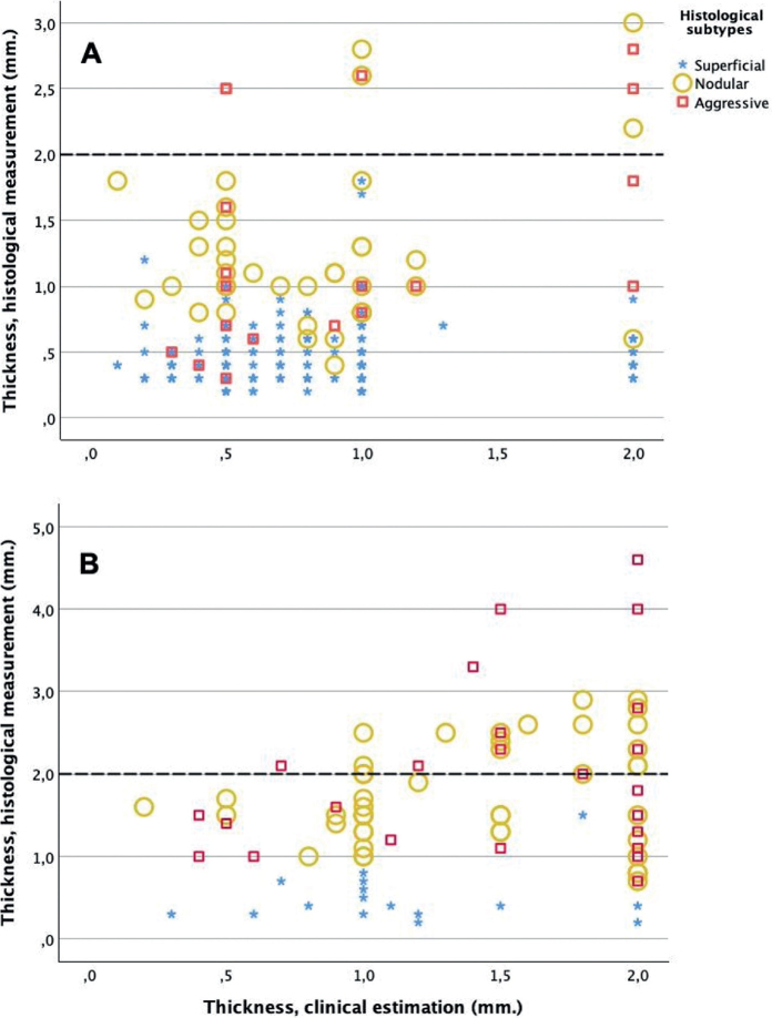
Fig. 3.
Scatterplots showing the corresponding clinical and histological measurements of basal cell carcinoma (BCC) thickness. The 3 histological tumour subtypes are distinguished with different markers. BCCs depicted in panel A were clinically evaluated as superficial, while those in panel B were clinically evaluated as nodular subtype. A dotted line at 2 mm is included to signify the thickness threshold pertinent to current recommendations for photodynamic therapy.

