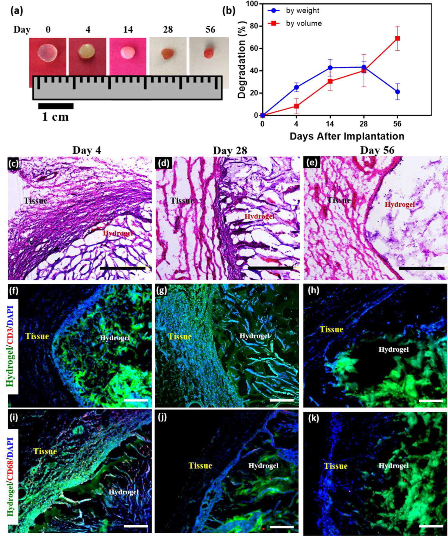
Figure 6.

In vivo biocompatibility and biodegradation of MeHA/ELP hybrid hydrogels using a rat subcutaneous implantation model. (a) Representative images of MeHA/ELP hydrogels before implantation (day 0) and on days 4, 14, 28, 56 post implantation. (b) In vivo biodegradation of MeHA/ELP hydrogels on days 0, 4, 14, 28, and 56 of implantation, based on weight and volume loss of the implants (n = 4). The in vivo degradation profile of MeHA/ELP hydrogels shows an approximately linear degradation behavior by volume during the first 56 days after implantation, as well as the highest biodegradation by weight between days 14 and 28. Hematoxylin and eosin (H&E) staining of MeHA/ELP sections (hydrogels with the surrounding tissue) after (c) 4, (d) 28, and (e) 56 days of implantation (scale bars = 500 μm). Immunohistofluorescent analysis of subcutaneously implanted MeHA/ELP hydrogels showing no significant local lymphocyte infiltration (CD3) at (f) 4, (g) 28, and (h) 56 days post implantation (scale bars = 200 μm). Fluorescent images showed transient macrophage infiltration (CD68) at (i) day 4, followed by no apparent positive fluorescence at (j) 28 and (k) 56 days post implantation (scale bars = 200 μm). Green, red and blue colors in (f–k) represent the MeHA/ELP autofluorescent hydrogels, the immune cells, and cell nuclei (DAPI), respectively. Hydrogels were formed by using 2% MeHA and 10% ELP at 120 s UV exposure time.
