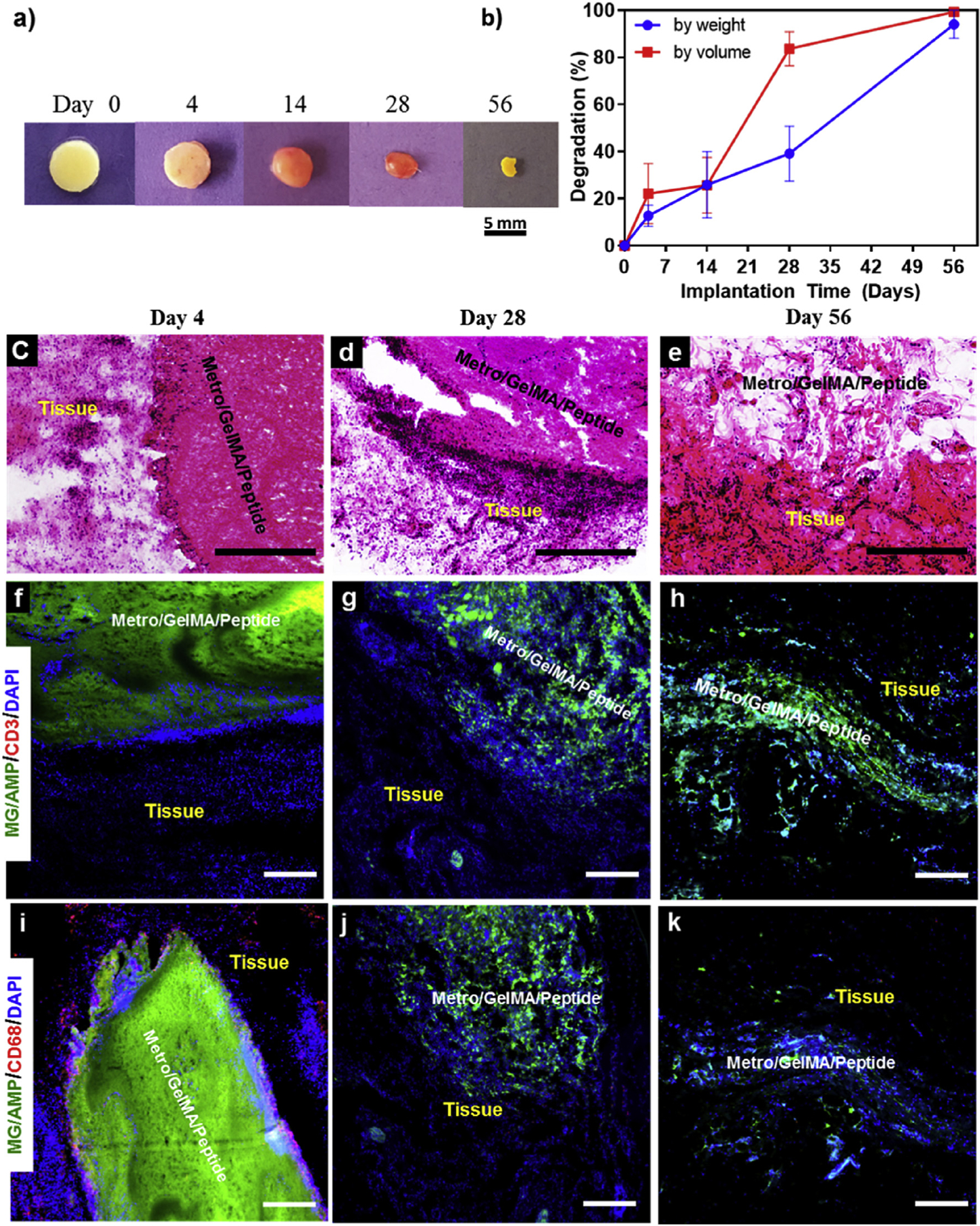
Fig. 7. In vivo biocompatibility and biodegradation of MeTro/GelMA-AMP composite hydrogel using a rat subcutaneous model.

(a) Representative images of the MeTro/GelMA-AMP hydrogels before implantation (Day 0) and on days 4, 14, 28, 56 post-implantation. (b) In vivo biodegradation of MeTro/GelMA-AMP hydrogels on days 0, 4, 14, 28 and 56 of implantation (n = 4). Hematoxylin and eosin (H&E) staining of MeTro/GelMA-AMP sections (hydrogels with the surrounding tissue) after (c) 4 days, (d) 28 days, and (e) 56 days of implantation (scale bars = 500 μm). (c) Fluorescent immunohistochemical analysis of subcutaneously implanted MeTro/GelMA-AMP hydrogels showing no significant local lymphocyte infiltration (CD3) at days (f) 4, (g) 28 and (h) 56 (scale bars = 200 μm), and exhibiting macrophages (CD68) at (i) day 4 but not at days (j) 28 and (k) 56 (scale bars = 200 μm). Green, red and blue colors in (f–k) represent the MeTro/GelMA-AMP hydrogels, the immune cells, and the cell nuclei (DAPI) respectively. 50/50 MeTro/GelMA hydrogels at 15% (w/v) total polymer concentration were used for the in vivo test.
