
Figure 1.
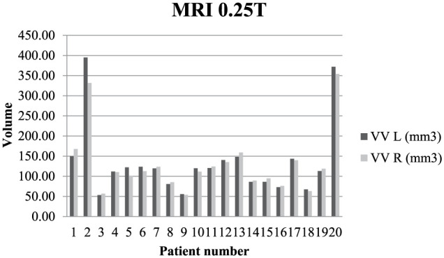
Variation in quantitative ventricular volume in 20 cats at 0.25 T. Note the mild asymmetry between the left and right ventricle in the first, second, fifth and sixth patients. Note the moderate ventriculomegaly in the second and 20th patients, and unilateral ventricular enlargement in fifth.
VV = ventricular volume; L = left; R = right