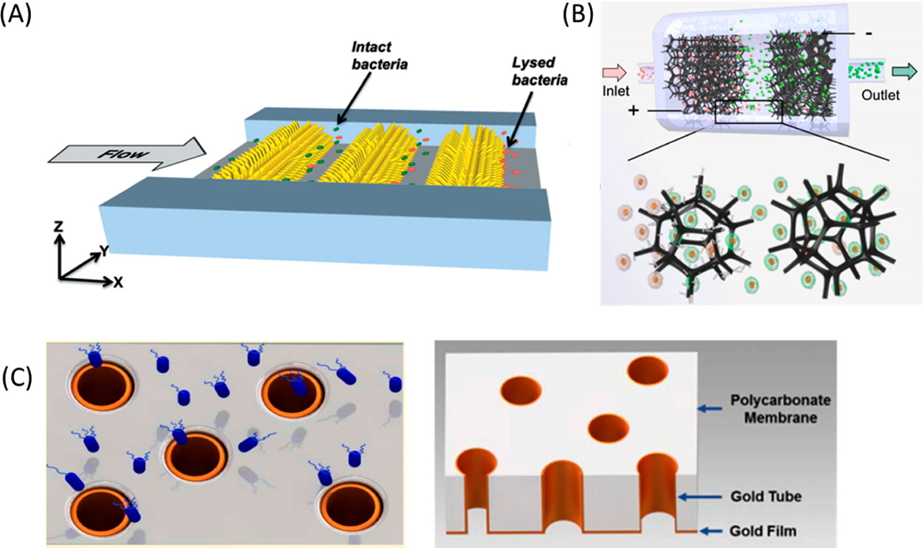
Figure 9.

Examples of enhanced electric fields at electrode edges or tips. (A) Illustration of cell lysis by three-dimensional sharp-tipped electrode (3DSTE) arrays in the microfluidic channel.236 Bacteria are lysed when they flow between nanoelectrodes. Reproduced with permission from ref 236. Copyright 2014 Royal Society of Chemistry. (B) Overview of the TENG-powered, nanowire-modified microfoam for continuous EP. Cell suspensions flowed through microfoam electrodes placed in parallel within the flow channel. Polypyrrole microfoam was modified by silver nanowires at the anode to enhance the electric field at the tip of the nanowire. The cathode microfoam was left unmodified to increase cell viability. Reproduced with permission from ref 237. Copyright 2020 Royal Society of Chemistry. (C) Schematic of E. coli (blue) flowing through gold microtube electrodes (left).246 Illustration showing 3D structure of gold microtube on the PC membrane (right). Reproduced with permission from ref 246. Copyright 2016 Royal Society of Chemistry.
