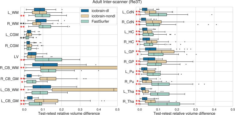
Figure 4.
The icobrain-dl measurements exhibited statistically significantly lower test-retest errors than icobrain-nondl and FastSurfer across all the subcortical structures (right) for adult cases (dataset 3.a) in inter-scanner settings, as quantified by relative test-retest volume differences. The asterisk colour indicates the better performing method (red = icobrain-dl, black = state-of-the-art). To ensure overall figure readability, certain boxplots have been cropped. L = left, R = right, WM = white matter, CGM = cortical gray matter, LV = lateral ventricles, CB = cerebellum, CdN = caudate nucleus, HC = hippocampus, GP = globus pallidus, Pu = putamen, Tha = thalamus.

