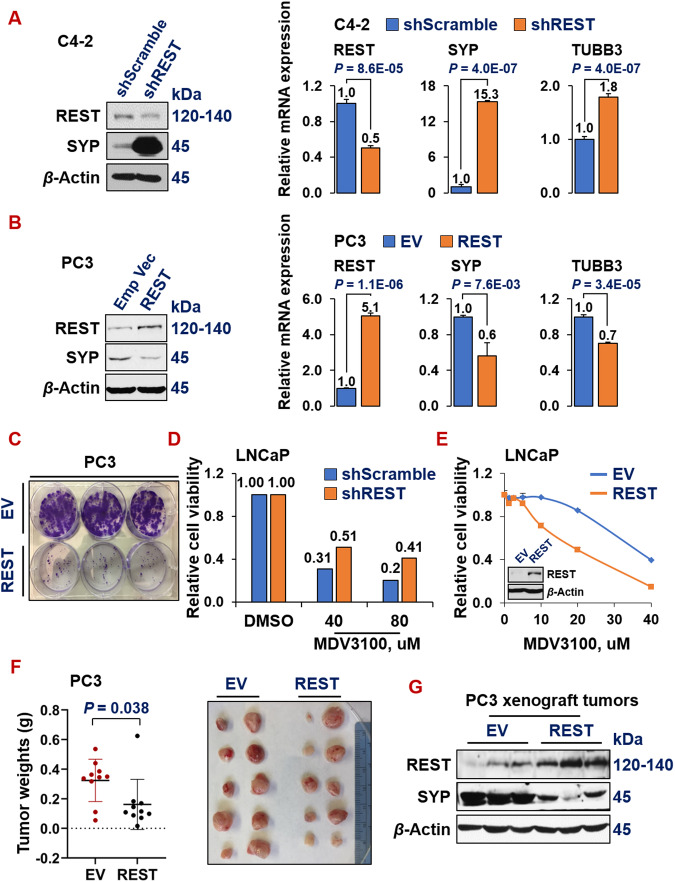
Fig. 2. REST inhibits NE marker expression, ADT resistance and tumor progression.
Western blotting and qPCR for REST and NE markers in NE-/low C4-2 cells upon REST silencing (A) and in NE + PC3 cells upon overexpressing REST cDNA (B). Y-axis shows relative fold changes in mRNA expression, normalized to beta actin. Error bars in PCR results represent standard deviation (s.d). Western blotting was carried on whole cell lysates from the indicated cell lines and for the indicated proteins. C Colony formation assay of PC3 cells carrying empty vector (EV) or REST cDNA. The two cell lines were seeded in triplicates in a 6-well plate, at 1000 cells/well and cultured for 14 days, followed by staining with crystal violet. D Viability and proliferation of LNCaP cells expressing either scramble shRNA or shREST, upon treatments of 40 µM or 80 µM of ADT drug MDV3100 for 3 days. E Cell viability and proliferation of LNCaP cells carrying either empty vector or REST cDNA, upon treating with a series doses of ADT drug MDV3100 for 3 days. Y-axis shows relative cell viability and proliferation, normalized to DMSO control. The experiment was carried out in quadruplicates each time, twice with similar results. Insert: Western blotting shows REST overexpression in PC3-REST cells. F Subcutaneous xenograft tumor growth of PC3 cells carrying either EV or REST cDNA in NOD/SCID male mice. Left: tumor weights at euthanization (P = 0.038) 8 weeks after implantation. Right: images of the xenograft tumors at euthanization (5 mice for each cell line, n = 10 tumors). G Western blotting for REST and NE marker SYP protein levels in xenograft tumors from PC3-EV and PC3-REST cells.

