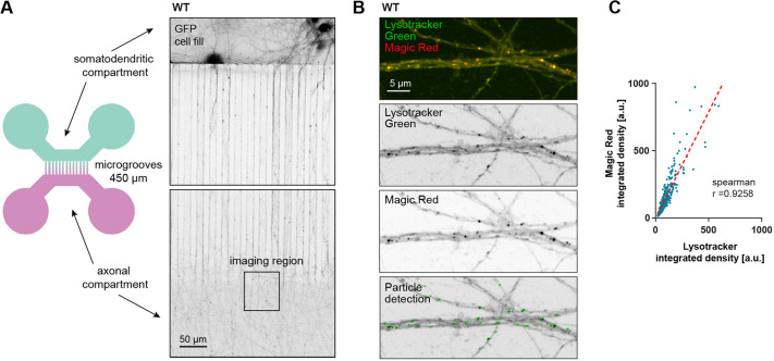
Fig. 3.
Lysotracker-positive vesicles in distal axons are degradatively active. (A) Illustration of the two-compartment MFC used for live-imaging of axonal trafficking (left panel), and microscopy image of DIV12 mouse hippocampal neurons expressing GFP (right panel). (B) Confocal image of axons inside MFC, labeled with Lysotracker Green and Magic Red. (C) Integrated density values of Lysotracker correlate with the integrated density of Magic Red; 815 particles were analyzed in three separate locations of one independent neuron culture in an area of ∼0.2 mm². The dotted red line indicates the calculated linear regression (y=1578×x+4156); Spearman's correlation coefficient, r=0.9258.

