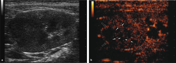
Figure 10.
Renal cell carcinoma in a cat. (a) B-mode ultrasound scan showing a hypoechoic lesion with internal echoes (open arrows), which could represent malignant tissue or alternatively debris/haemorrhage in a benign lesion. (b) On CEUS, there is uptake of contrast in the arterial phase, with the presence of feeding vessels (small arrows) indicating malignancy

