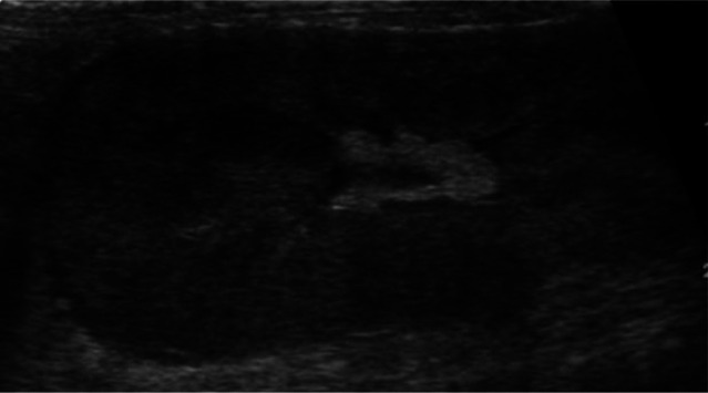
Figure 4.
Sagittal ultrasound scan of the left kidney of a cat with feline infectious peritonitis showing renomegaly (5.1 cm), which was bilateral, and an irregular contour. The kidney is markedly increased in echogenicity and there is poor corticomedullary distinction. A thin, crescent-shaped, hypoechoic subcapsular rim is present

