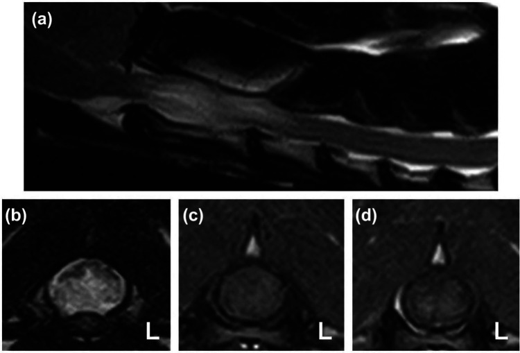
Figure 1.
T2-weighted (T2W) sagittal (a) and transverse (b) images of the cranial cervical spinal cord of cat 1 showing marked spinal cord swelling at the level of C2. The transverse T2W image reveals the ventral half of the spinal cord to be diffusely hyperintense with a relatively sharp distinction to the dorsal spinal cord. T1-weighted pre-contrast (c) and post-contrast (d) transverse images at the level of C2 reveal faint contrast enhancement in the ventral spinal cord predominantly affecting the gray matter

