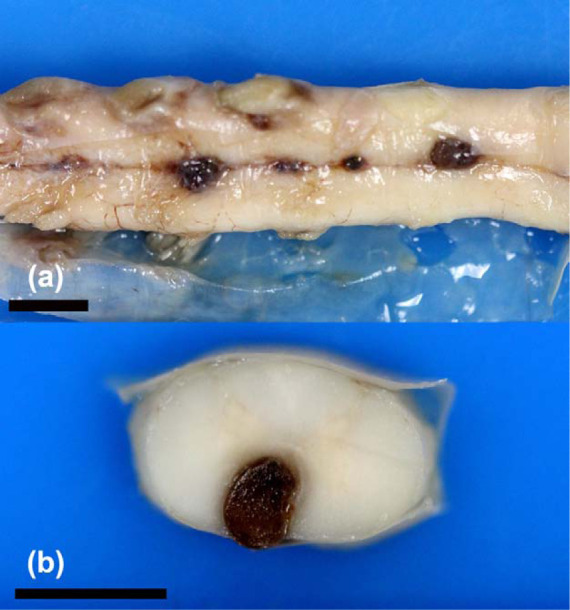
Figure 4.

(a) Several dark raised nodules are evident on the ventral aspect of the cervical spinal cord in cat 4 from C3 to C5 at the level of the ventral spinal artery, consistent with multifocal vascular ectasia (aneurysm). Formalin-fixed tissue. (b) Cross-section of the cervical spinal cord. The nodule has compressed the regional ventral spinal cord parenchyma. Formalin-fixed tissue. Bars = 5 mm
