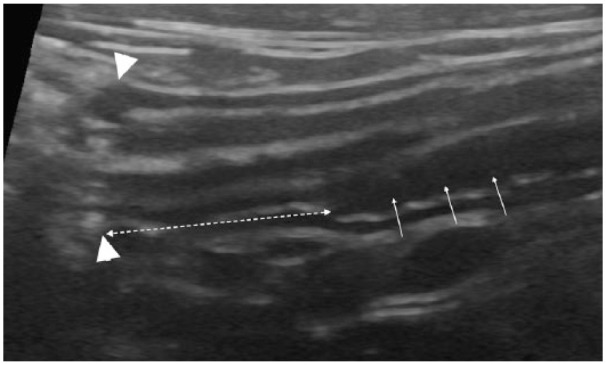
Figure 3.

Longitudinal ultrasonographical image of the ileum and ileocolic junction in a clinically healthy, young cat. An asymmetrically positioned hypoechoic extra layer (APHEL) is present in the deepest part of the intestinal wall, in the ileum (white arrows). The ileocolic junction is to the left in the image (arrowheads). The dashed double arrow shows the distance between the ileocolic junction and the most distal margin of the APHEL
