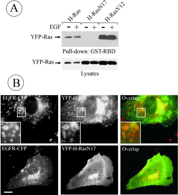
Figure 4.
Colocalization of dominant-negative H-RasN17 with endosomal EGFRs. (A) Cells were transiently transfected with YFP-H-Ras, YFP-H-RasN17, or YFP-H-RasV12; lysed; and the GTP-bound Ras proteins were affinity precipitated (pulled down) from the lysates by using GST-RBD beads. Isolated proteins and aliquots of lysates were separated by electrophoresis, transferred to nitrocellulose membrane, and YFP-fused proteins were detected by blotting with GFP antibodies. (B) PAE/EGFR-CFP cells were transiently transfected with wild-type YFP-H-Ras or YFP-H-RasN17. Living cells were imaged through CFP and YFP filter channels. Insets, high-magnification images of the regions indicated by white rectangles. Bar, 5 μm.

