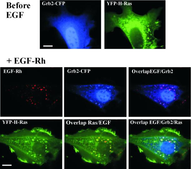
Figure 5.
Colocalization of Grb2-CFP and YFP-H-Ras in NIH 3T3 cells. NIH 3T3/EGFR cells were transiently transfected with Grb2-CFP and YFP-H-Ras. Live-cell YFP and CFP images were acquired before and after incubation of cells with 200 ng/ml EGF-Rh for 20 min at 37°C. EGF-Rh was detected using Cy3 filter channel. Bar, 10 μm.

