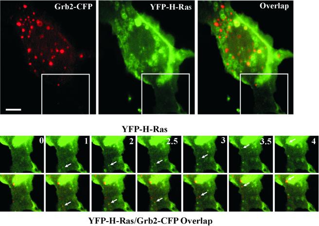
Figure 6.
Visualization of the formation and movement of endocytic vesicles containing YFP-H-Ras and Grb2-CFP. PAE/EGFR cells transfected with Grb2-CFP and YFP-H-Ras were grown in microscope chambers. Serum-starved cells were incubated with 100 ng/ml EGF for 5 min at 37°C, and 20 images were acquired with 30-s intervals at 37°C (time is indicated in minutes). Large images are taken at time 0. Selected time-lapse YFP and YFP/CFP overlapped images of the region of the cell indicated by the white rectangle are presented at the bottom. Arrows point to the vesicle containing both Grb2-CFP and YFP-H-Ras that appears at time 1 min and moves toward the perinuclear area of the cell. Quick time movie is available in the supplemental materials. Bar, 5 μm.

