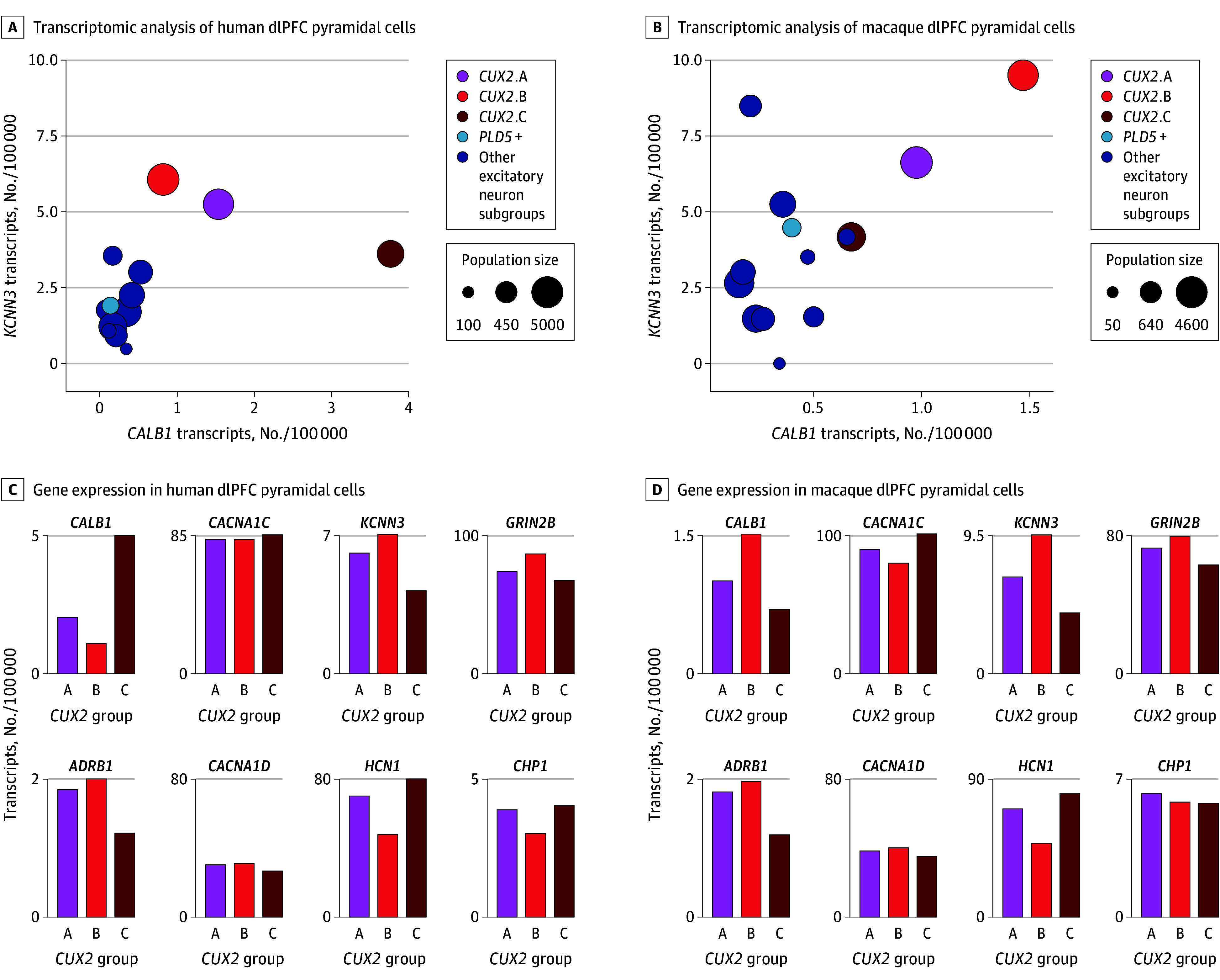
Figure 1. Transcriptomic Analyses of Excitatory Neurons in the Dorsolateral Prefrontal Cortex (dlPFC) of Humans and Macaques.

A, Transcriptomic analyses of human dlPFC pyramidal cells, showing expression levels of CALB1 (encoding the calcium-binding protein calbindin) and KCNN3 (encoding the SK potassium channel opened by calcium, SK3). Population sizes are expressed as number of nuclei out of 20 000. The CUX2-expressing cells have higher CALB1 than other excitatory cells and very high levels of calcium-related genes, including KCNN3. B, Bar graph quantification of CALB1, CACNA1C, KCNN3, GRIN2B (encoding the GluN2B subunit of the NMDA receptor that fluxes high levels of calcium), ADRB1 (encoding the β1-adrenoceptor [β1-AR], which drives Cav1.2 actions in the heart during stress exposure), CACNA1D (encoding the LTCC Cav1.3), HCN1 (encoding the hyperpolarization-activated and cyclic nucleotide–gated channel opened by cyclic adenosine monophosphate), and CHP1 (encoding the calcineurin inhibitor calcineurin homologous protein 1), in the 3 CUX2 (CUX2 A-C) dlPFC populations in the human dlPFC. C, Same as panel A but in the macaque dlPFC. D, Same as panel B but in the macaque dlPFC. Note lower levels of CACNA1D vs CACNA1C in both species. Expression values are normalized counts of the number of transcripts per 100 000 in each cell type.
