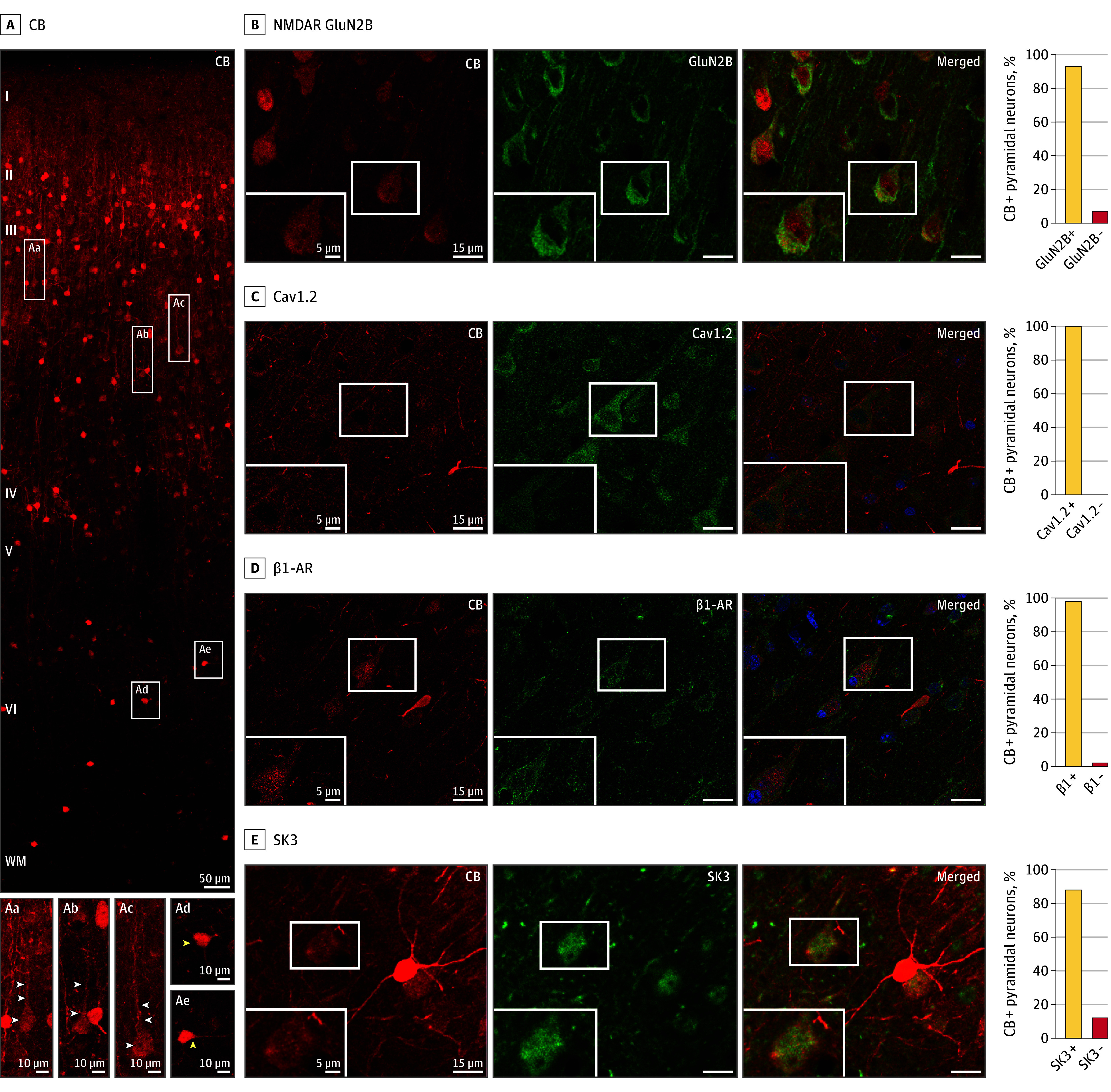
Figure 2. Calbindin-Expressing Pyramidal Cells in Layer III of the Dorsolateral Prefrontal Cortex (dlPFC) of Macaques and NMDA Receptor–GluN2B, Cav1.2 Channels, SK3 Channels, and β1-Adrenoceptor (β1-AR).

A, Expression of the calcium-binding protein calbindin (CB), across the cortical column in the dlPFC of macaques. Pyramidal cells (examples in white rectangles) are concentrated in layer III and have modest calbindin expression (Aa-Ac; white arrowheads), while interneurons are throughout all layers, especially in layer II, and have intense calbindin expression (Ad-Ae; yellow arrowheads). B-E, Calbindin-expressing pyramidal cells coexpress the following proteins: B, NMDA receptor (NMDAR) GluN2B; C, Cav1.2 (amplified with biotin-streptavidin); D, β1-adrenoceptor (β1-AR) (amplified with biotin-streptavidin); and E, SK3. The intensely labeled, bright red neurons (eg, in E) are calbindin-expressing GABAergic interneurons, which have higher levels of calbindin than pyramidal cells. The percentage of calbindin-expressing pyramidal cells coexpressing each protein is shown on the right. Where present, blue labeling is the Hoechst nuclear counterstain.
