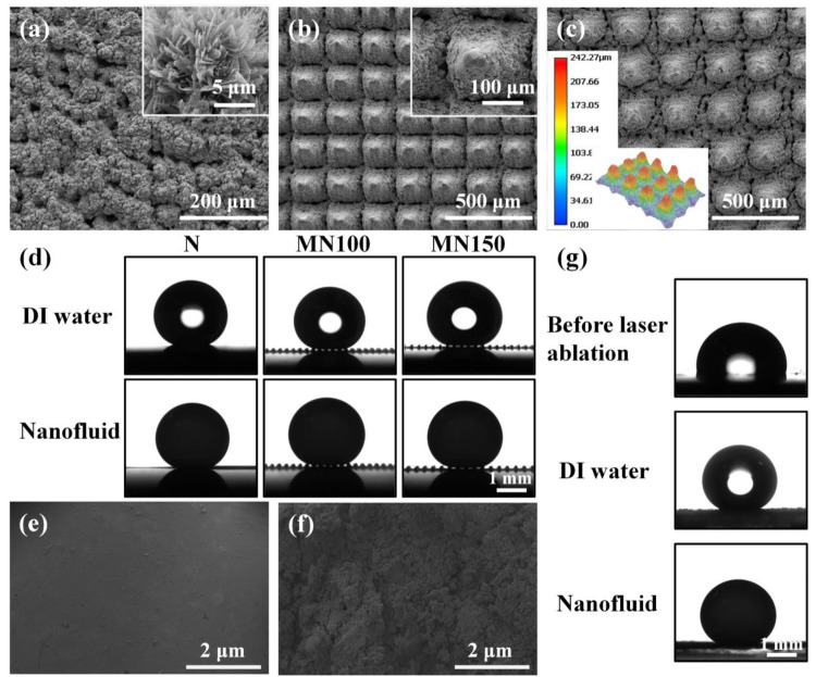
Figure 2.
SEM images of (a) N superhydrophobic Cu surface, (b) MN100 superhydrophobic Cu surface, and (c) MN150 superhydrophobic Cu surface, respectively. (d) Local DI water contact angle and local nanofluid contact angle on the N, MN100, and MN150 superhydrophobic Cu surfaces, respectively. SEM images of PDMS elastic surface (e) before laser ablation and (f) after laser ablation. (g) The local DI water contact angle on the elastic PDMS surface before laser ablation, DI water contact angle after laser ablation, and nanofluid contact angle after ablation, respectively.

