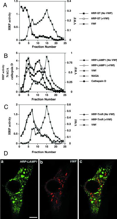
Figure 4.
vWF does not affect the localization of HRP-ST, HRP-LAMP1, and HRP-TrnR in AtT-20 cells. (A–C) Cells transiently expressing either HRP-ST, HRP-LAMP1, or HRP-TrnR in the absence or presence of vWF expression were fractionated on 0.7–1.75 M sucrose equilibrium gradients followed by measurement of HRP activity across the gradient (see MATERIALS AND METHODS). The amounts of vWF (∗), NAGA activity (+), and cathepsin D (♦) across the gradient were measured as described in MATERIALS AND METHODS. (A) Distribution of ST-HRP expressed on its own (▪) or coexpressed with vWF (□). (B) Distribution of HRP-LAMP1 expressed on its own (●) or coexpressed with vWF (○). (C) Distribution of HRP-TrnR expressed on its own (▴) or coexpressed with vWF (▵). (D) Immunofluorescence labeling of cells transiently coexpressing HRP-LAMP1 and vWF. Cells were fixed and permeabilized as described in MATERIALS AND METHODS and then colabeled with rabbit polyclonal anti-HRP (a) and sheep polyclonal anti-vWF (b). A merger of two composite channels is shown in c. Bar, 5 μm.

