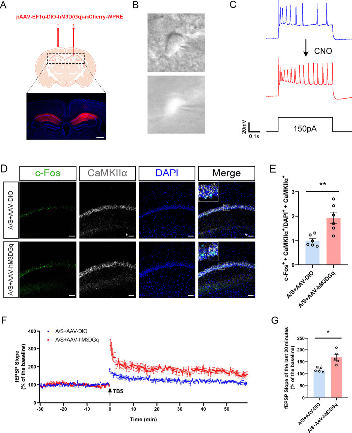
Fig. 9.
Effect of specific activation of hippocampal CA1 glutamatergic neurons on synaptic plasticity. A Fluorescence images showed that the chemogenetics vector was effectively expressed in the CA1 region of the hippocampus. B The schematic diagram of glutamatergic neurons in the hippocampal CA1 region recorded under microscope in acute brain sections. C In vitro infusion of CNO (10 mM) increased the firing frequency of glutamatergic neurons in the hippocampal CA1 region of AAV-hM3D(Gq)-injected POCD mice. D Representative colabeling images of c-Fos and CaMKIIα. Scale bar, 100 μm. E Quantitation of the co-labeling rate of c-Fos and CaMKIIα **p < 0.01 (n = 6). F LTP recorded in the CA1 region of the hippocampus. The arrow indicates the point in time of the TBS. G Average fEPSP slope during the last 20 min after TBS *p < 0.05 (n = 5)

