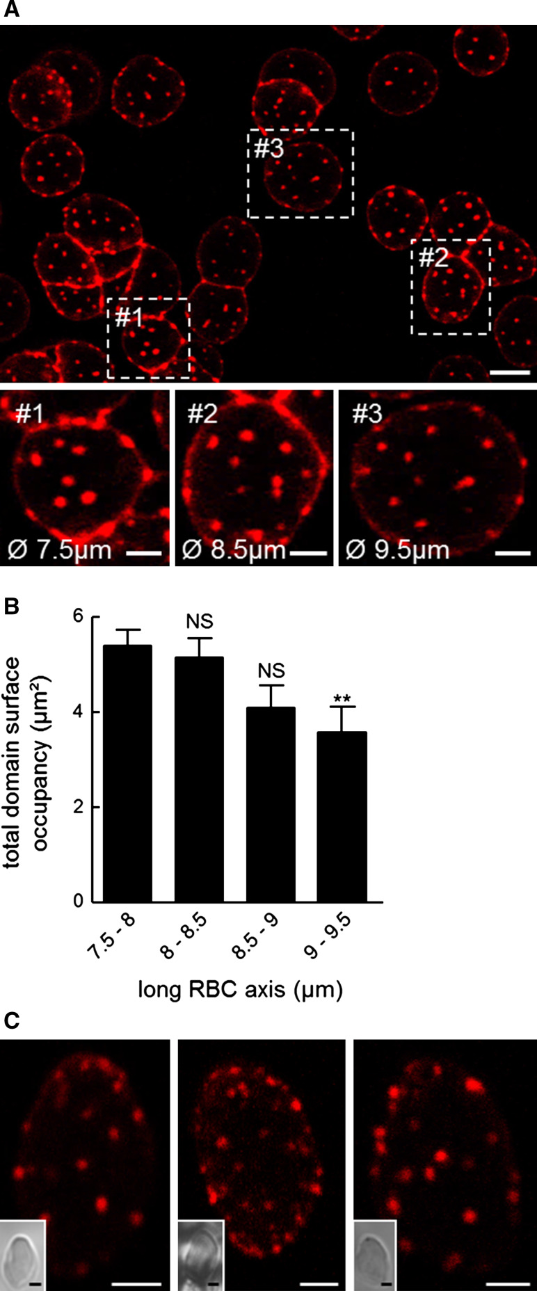
Fig. 5.
Cholesterol submicrometric domains largely resist membrane stretching and are similarly abundant in gel-suspended (3D), non-stretched RBCs. A Confocal imaging of domains on differentially spread RBCs. RBCs were labeled with theta*, washed, attached-spread and visualized at 20 °C. RBC attachment on poly-l-lysine leads to variable stages of RBC spreading (boxed areas #1 to #3 are enlarged at insets). Theta*-enriched domains resist membrane stretching but decrease in size. Scale bars 5 µm at general view and 2 µm at insets. B Morphometry of domain surface occupancy. Surface of theta*-labeled domains was determined per hemi-cell surface and values were classified according to RBC long diameter. Mean ± SEM of total domain area recorded on 8–26 cells from two independent experiments. Kolmogorov–Smirnov test was performed using the 7.5–8 µm class as reference; NS not significant, **p < 0.01. C Confocal imaging of domains on RBCs trapped in a 3D-gel. RBCs were labeled with theta*, washed and trapped as suspension in hydrogel, then directly observed by confocal microscopy at 37 °C. Insets show preservation of RBC ovoid shape. Images representative of six independent experiments. Scale bars 2 µm

