Fig. 9.
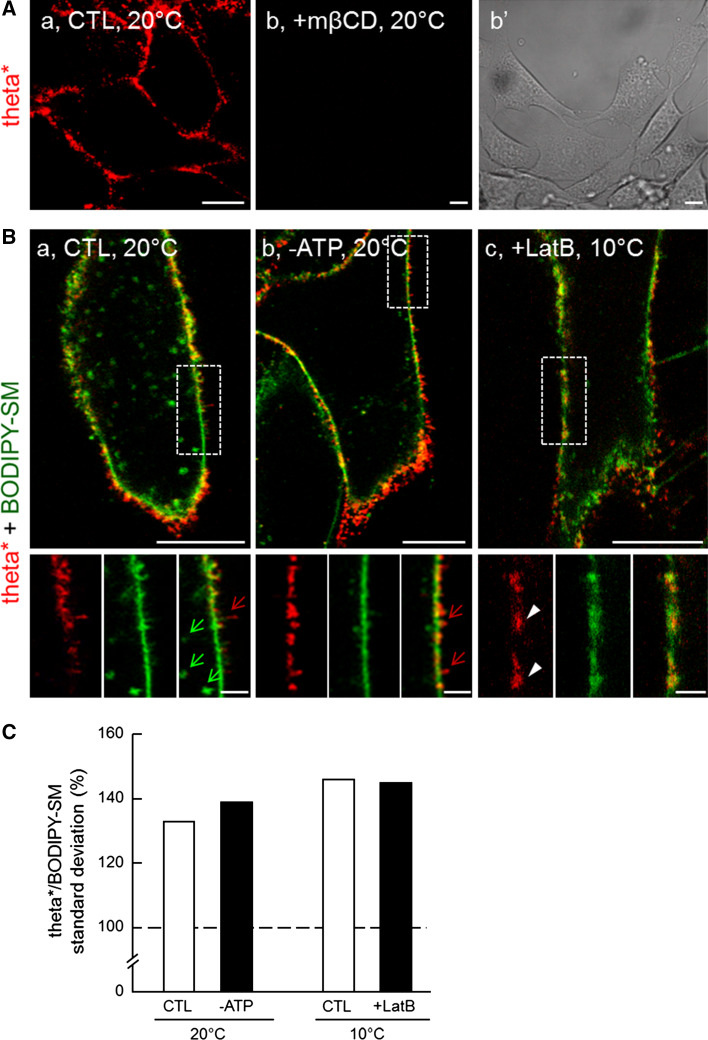
Theta* labels submicrometric domains at the lateral plasma membrane of C2C12 myoblasts. A Theta* labels clusters sensitive to cholesterol depletion. C2C12 myoblasts in IBIDI chambers were pre-incubated or not (a) with 5 mM mβCD at 37 °C (b, b′), then labeled with theta* and visualized by confocal imaging at 20 °C. b′ shows presence of myoblasts in b. B Cholesterol heterogeneity does not primarily reflect endocytotic structures nor ruffles. C2C12 were pre-incubated (c) or not (a, b) with latrunculin B (LatB; to inhibit actin polymerization), then labeled with theta* and BODIPY-SM in presence (b) or not (a, c) of sodium azide and deoxyglucose (-ATP; to prevent endocytosis by energy depletion) and visualized at 20 °C (a, b) or 10 °C (to prevent endocytosis by low temperature, c). Green and red arrows indicate deep endocytotic structures and membrane ruffles respectively; white arrowheads point to cholesterol-enriched lateral domains. Scale bars 10 µm at general views and 2 µm in insets. C Quantification. Mean intensity and associated standard deviations (SDs) were quantified for both theta* and BODIPY-SM signals. SDs were then normalized as percentage of the corresponding mean intensity to yield variation coefficients that were further presented as ratios between theta* and BODIPY-SM. Results are mean of two to three independent experiments pooled from 29 to 50 area per condition. Dotted line indicates theoretical level of equal variation coefficient between theta* and BODIPY-SM

