Fig. 4.
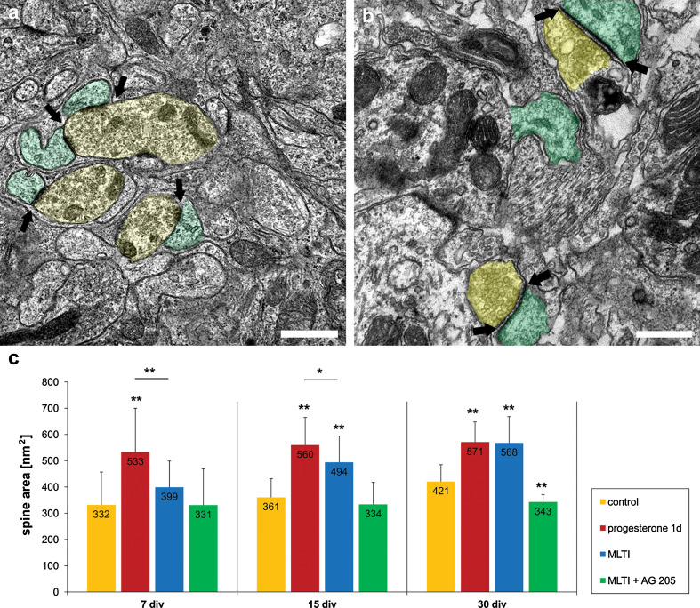
Electron microscopic analysis of spine area. a, b Electron microscopic analysis of the dendritic spines revealed numerous chemical synapses, with synaptic vesicles inside. Presynaptic component with synaptic vesicles is shaded in yellow, postsynaptic component is shaded in green; arrows indicate the postsynaptic density. a Dendritic spines and synapses in controls after 15 days in vitro. b Spines and synapses after 24 h progesterone treatment in PC after 15 div. The area of dendritic spines and postsynaptic density are increased compared to controls. c Size of the dendritic spines measured in PC after 7, 15, and 30 div in controls and after progesterone incubation for 24 h, (mifepristone long-time incubation, MLTI) and MLTI plus AG 205; error bars SD. Scale bars (a, b) 1 μm

