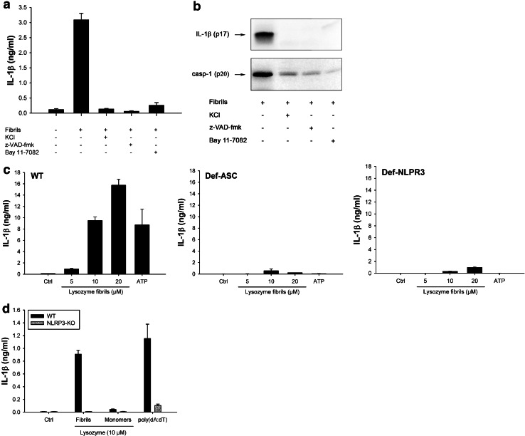
Fig. 3.
Lysozyme fibrils activate the NLRP3 inflammasome. a, b PMA-primed THP1 cells were incubated for 6 h with 5 μM lysozyme fibrils in the presence or absence of either KCl (130 nM), the pan-caspase inhibitor z-VAD-fmk (20 μM), or the NLRP3 inhibitor Bay 11-7082 (20 μM). Cell supernatants were analyzed for IL-1β by ELISA (a) and immunoblotting (b, upper panel) or for activated caspase-1 (casp-1) (b, lower panel); c PMA-primed WT, ASC-deficient (Def-ASC) and NLRP3-deficient (Def-NLRP3) THP1 cells were incubated for 6 h with ATP (2 mM) or the indicated amounts of lysozyme fibrils, and IL-1β was quantified by ELISA in the cell supernatant; d LPS-primed BMDMs from WT (black bars) or NLRP3 KO (grey bars) mice were stimulated for 21 h with the indicated amounts of either lysozyme fibrils or monomers, or transfected with poly(dA:dT) (5 μg/ml), and cell supernatants were analyzed for IL-1β by ELISA. n = 3, mean ± SD

