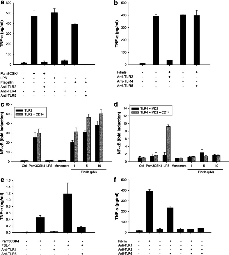
Fig. 5.
Lysozyme fibrils activate the TLR2/TLR1 heterodimer. a, b THP1 cells were incubated for 6 h with 1 ng/ml Pam3CSK4, 5 ng/ml LPS, 100 ng/ml flagellin (a) or with 5 μM lysozyme fibrils (b), in the presence or absence of 20 μg/ml anti-TLR2, anti-TLR4, or anti-TLR5 antibodies. TNF-α was quantified in the cell supernatant by ELISA; c, d HEK 293 cells expressing TLR2 (c) or TLR4 and MD2 (d) with or without CD14 (grey and black bars, respectively) were incubated 6 h in the presence of Pam3CSK4 (10 ng/μl), LPS (100 ng/μl), or the indicated concentrations of either lysozyme monomers or fibrils. NF-κB activation was detected by quantifying luciferase activity in cell lysates; e, f THP1 cells were incubated for 6 h with 1 ng/ml Pam3CSK4, 100 ng/ml FSL-1, or with 5 μM lysozyme fibrils, in the presence or absence of 20 μg/ml anti-TLR2, anti-TLR1, or anti-TLR6 antibodies. TNF-α was quantified in the cell supernatants by ELISA. n = 3, mean ± SD

