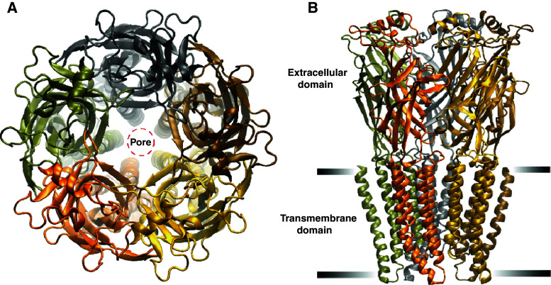
Fig. 1.
a A top view schematic of a pLGIC showing its five-fold symmetry and the central ion permeation pathway. The five M2 α-helical segments, located within transmembrane domain, line a pore. Each subunit is given a different colour. b A side view schematic of a pLGIC showing two of the three functional domains, the extracellular and transmembrane (demarcated by black horizontal bars). The intracellular domain is not shown. The images were made using the crystal structure of the C. elegans α GluClR 3RIF [5]

