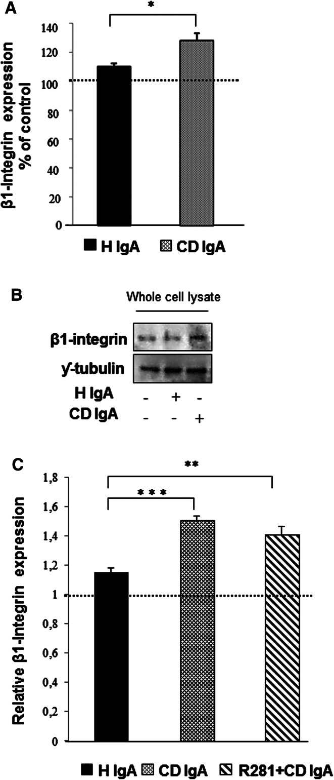
Fig. 5.
Celiac patient immunoglobulins A (CD IgA) increase β1-integrin expression in endothelial cells after 24-h treatment. a Quantification of Western-blot analyses of β1-integrin in whole endothelial cell lysates under different experimental conditions. Protein expression was normalized to γ-tubulin and control was taken as 100 %. The dashed line indicates HUVECs without any treatment. Bars represent mean of data derived from four independent experiments repeated in duplicate and error bars indicate standard error of the mean. A p value <0.05 was considered significant (*p < 0.05) and only statistically significant results are reported. b Representative Western blot of β1-integrin in whole endothelial cell lysates under different experimental conditions. c The relative amount of β1-integrin was analyzed by microtiter plate method in unpermeabilized HUVECs alone or incubated with CD IgA and healthy (H IgA) subject’s immunoglobulins. The dashed line indicates HUVECs without any treatment. Bars signify mean and error bars standard error of the mean. A p value <0.05 was considered significant (**p < 0.01, ***p < 0.001) and only statistically significant results are reported. Data are derived from three independent experiments repeated in quadruplicate. The extracellular transglutaminase 2 activity was inhibited by administering a site-directed, non-permeable inhibitor, R281

