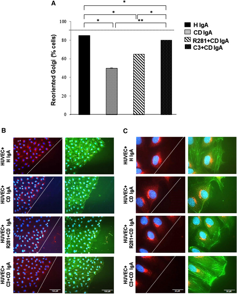
Fig. 7.
Celiac patient immunoglobulins A (CD IgA) inhibit endothelial cell polarization and migration. a Quantification of cell polarization in HUVECs on fibronectin incubated with CD IgA and healthy (H IgA) subject’s immunoglobulins after 24-h treatment by Golgi-reorientation assay. Bars signify the percentage of HUVECs which had reoriented Golgi towards the wound edge and error bars indicate standard error of the mean. A p value <0.05 was considered significant (*p < 0.01, **p < 0.001) and only statistically significant results are reported. More than 500 cells were selected over three independent experiments. b, c Representative immunofluorescence stainings of Golgi by B-COP antibody (red) in HUVECs under different experimental conditions. Actin cytoskeleton was visualized by phalloidin (green) and DAPI (blue) was used as nuclear counterstaining. Images were selected over three independent experiments. The white dashed lines represent the edge of the wound. b Scale bar 100 μM and c scale bar 20 μM. The extracellular TG2 activity was inhibited by administering a site-directed non-permeable inhibitor, R281. Similarly, C3 transferase was administered to inhibit small Rho GTPases in the phenotype

