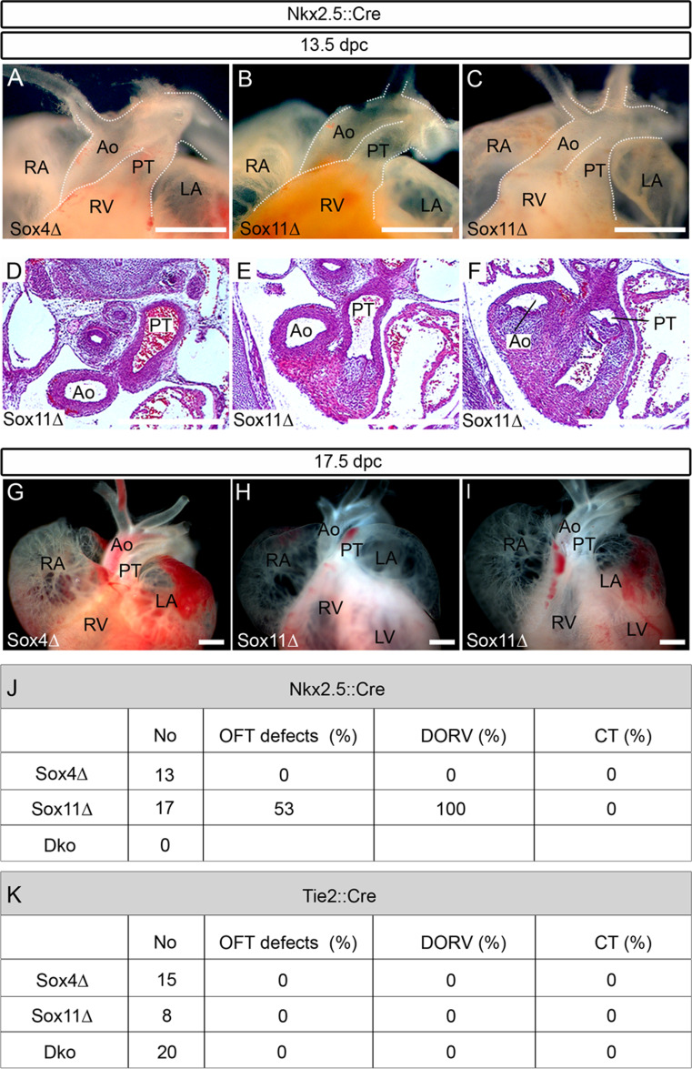
Fig. 4.
Developmental cardiac outflow tract defects in mice with mesodermal or endothelial cell-specific SoxC gene ablations. Macroscopic appearance of the anterior heart pole (a–c, g–i) and hematoxylin-eosin stainings of corresponding consecutive serial sections (horizontal plane) (d–f) from embryos with Nkx2.5::Cre-mediated, mesodermal SoxC gene ablations at 13.5 dpc (a–f) and 17.5 dpc (g–i). Analyzed genotypes included: Sox4 ΔNkx2.5 (Sox4Δ) (a, g) and Sox11 ΔNkx2.5 (Sox11Δ) (b–f, h, i). For age-matched wild-type controls, see Figs. 1 and 2. DORV was observed in a fraction of Sox11 ΔNkx2.5 embryos (c–f, i). Ao aorta, LA left atrium, RA right atrium, RV right ventricle, PT pulmonary trunk. Scale bars 500 μm. j Overview of the number of animals obtained with mesodermal Nkx2.5::Cre-mediated SoxC deletions, and the percentage of outflow tract defects (OFT defects) per genotype in total and subdivided into DORV and CT. k Summary of the number of animals obtained with Tie2::Cre-mediated, endothelial SoxC deletions, and the percentage of outflow tract defects per genotype in total, and subdivided into DORV and CT

