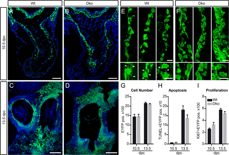
Fig. 6.
Analysis of general properties and morphology of cardiac neural crest in mice with neural crest-specific SoxC gene ablations. a–d Immunohistochemistry for EYFP (green) was performed at 10.5 dpc (a, b) and 13.5 dpc (c, d) on embryos carrying a combination of Rosa26 stopflox-EYFP and Wnt1::Cre alleles (Wt) (a, c) or carrying the Rosa26 stopflox-EYFP on a Sox4 ΔWnt1 Sox11 ΔWnt1 background (Dko) (b, d). Nuclei are counterstained with DAPI (blue). e, f High-magnification confocal images of cardiac neural crest cells (green) at 10.5 dpc taken from the wild-type (e) or age-matched Sox4 ΔWnt1 Sox11 ΔWnt1 (f) embryos. Wnt1::Cre-dependent activation of EYFP from the Rosa26 stopflox-EYFP allele was used to label cells of the neural crest lineage. Lower panels show even higher magnification of single cells shown in e, f. Arrowheads mark filopodia between neural crest cells. Scale bars 100 μm (a–d), 15 μm (e, f). g–i Quantification of the total number of cardiac neural crest cells per section (g), the TUNEL-positive apoptotic fraction (h), and the Ki67-positive proliferating fraction (i) at 10.5 and 13.5 dpc in outflow tract regions of embryos carrying a combination of Rosa26 stopflox-EYFP and Wnt1::Cre alleles (Wt, black bars) or carrying the Rosa26 stopflox-EYFP on a Sox4 ΔWnt1 Sox11 ΔWnt1 background (Dko, grey bars). At least four separate sections from three independent embryos were counted for each genotype. Data are presented as mean ± SEM. No statistically significant differences were observed between the two genotypes

