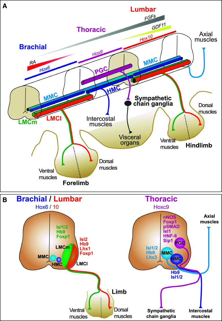
Fig. 1.
Topographic organization and molecular markers of motor columns along the anteroposterior axis of the developing spinal cord. a Schematic diagram illustrating the three-dimensional distribution of the motor columns. Graded levels of the morphogens FGFs, RA, and GDF11 determine spatial expression of Hox paralog groups that shape the spinal cord into a brachial, a thoracic, and a lumbar portion. At thoracic levels, MNs are distributed in a medial motor column (MMC) that innervates the axial muscles, a hypaxial motor column (HMC) connected to the intercostal muscles and a column of preganglionic MNs (PGC) that project to the sympathetic ganglia chain, which innervates visceral organs. At brachial and lumbar levels, MNs gather in an MMC column, a restricted HMC column, and a lateral motor column (LMC) that innervates locomotor muscles. MNs of the LMC are divided into a medial (LMCm) and a lateral (LMCl) complement that innervate ventral or dorsal limb muscles, respectively. b Location of each motor column and their molecular markers on schematic transverse sections in embryonic spinal cord at brachial/lumbar or at thoracic levels. Hox6 are determinants of the brachial MNs and Hox10 are required for lumbar MN fate, while Hoxc9 establishes thoracic MN identity. FGFs fibroblast growth factors, RA retinoic acid, GDF11 growth differentiation factor 11

