Fig. 1.

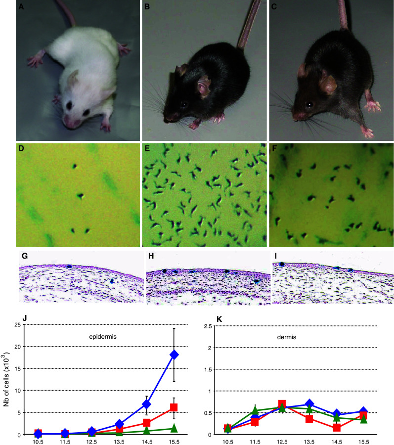
Melanoblast development in wt mice and β-catenin coat color mutants. a–c β-catenin loss-of-function (Δbcat), wild-type (wt), and gain-of-function (bcat*) adult mice. (A, D, G: Δbcat), (B, E, H: wt) and (C, F, I: bcat*). Note that melanocyte-specific disruption of β-catenin (Δbcat) led to the complete absence of pigmentation, whereas a stabilized and nuclear form of β-catenin (bcat*) in melanocytes leads to a less intense coat color than controls (wt). d–f Macroscopic observations of the trunk region of Δbcat, wt, and bcat* E14.5 embryos. The melanoblasts, identified as X-gal-positive cells, are less abundant in both β-catenin mutants than in wt, and in Δbcat than in bcat*. g–i Sections through the trunk of embryos at E13.5. Melanoblasts, the X-gal positive cells, are observed in both compartments of the skin: epidermis (ep) and dermis (de). j–k The total number of epidermal and dermal melanoblasts in the trunk was estimated from E10.5 onwards. In the epidermis (j), the number of melanoblasts increased with time with the most substantial increase in wt mice and the smallest in Δbcat mice. In the dermis (k), the total number of melanoblasts remained low for all genotypes and was fairly constant from E11.5 onwards
