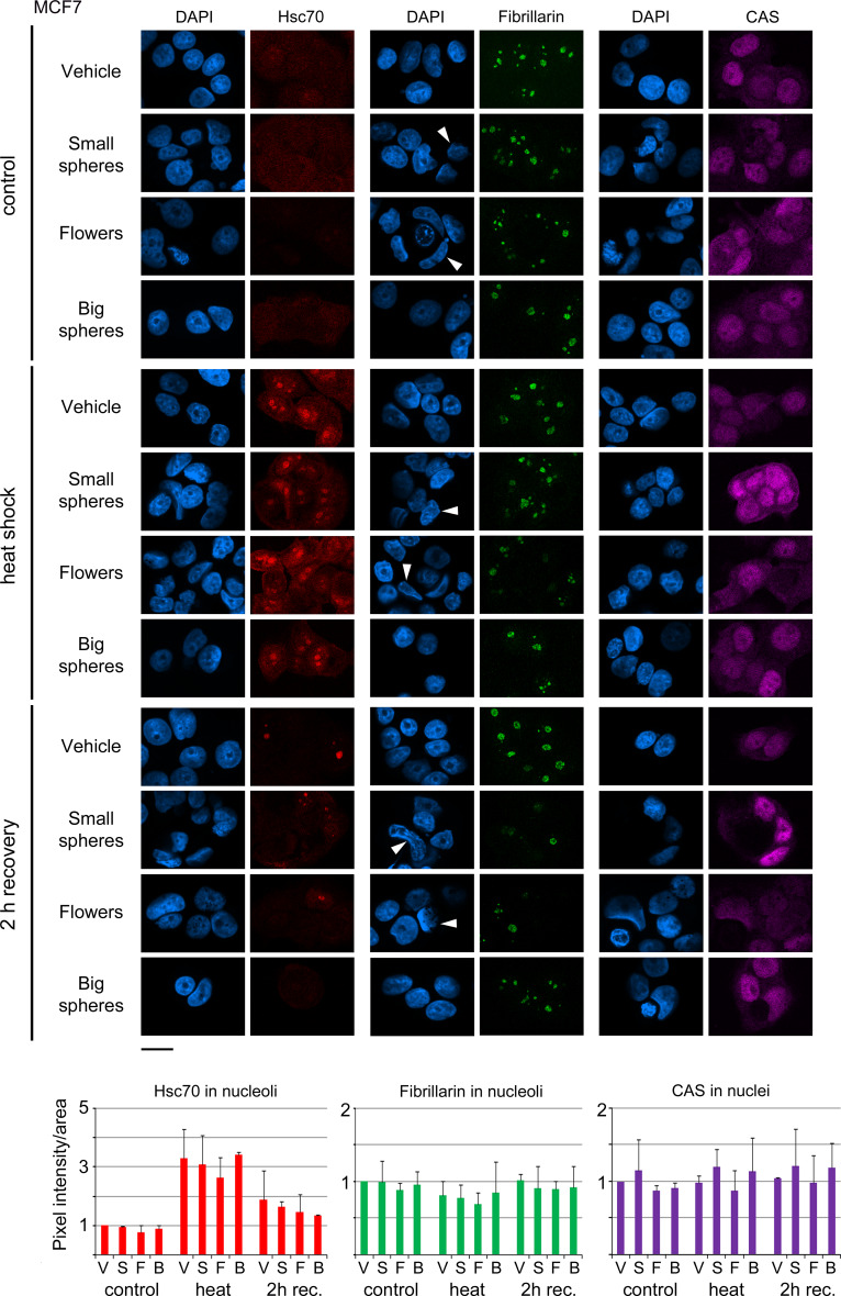
Fig. 2.
Impact of GNPs on the nuclear and nucleolar organization of cancer cells. MCF7 cells incubated with vehicle, small gold nanospheres (small spheres), nanoflowers (flowers), or big gold nanospheres (big spheres) were analyzed in the absence of heat (control), after heat shock, or 2 h recovery from heat stress. Hsc70, fibrillarin, or CAS were located by indirect immunofluorescence and images were acquired by confocal microscopy with identical settings for each antigen. Nucleolar fluorescence was quantified following established protocols [45]. Nuclei were stained with DAPI; size bar is 20 μm. Several of the irregularly shaped nuclei in GNP-treated samples are marked by arrowheads. Pixel intensities/area were measured for at least 66 nucleoli (a minimum of 21 cells) for each data point and experiment. Bar graphs depict the average + STDEV of two independent experiments. V vehicle, S small gold nanospheres, F gold nanoflowers, B big gold nanospheres. Results are normalized to controls which were incubated with vehicle alone

