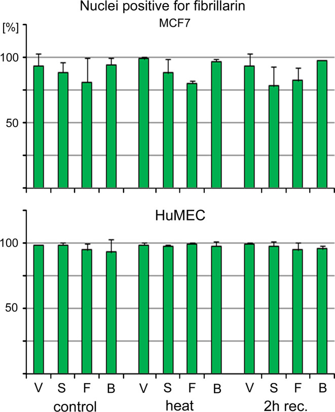
Fig. 4.
Staining of MCF7 and HuMEC nuclei with antibodies against fibrillarin. The percentage of nuclei that are stained with antibodies against fibrillarin was determined for MCF7 cells and HuMEC. Samples were treated as described for Figs. 2 and 3, and nuclei positive for fibrillarin staining were assessed in two independent experiments. Original data are shown as average + STDEV. V vehicle, S small gold nanospheres, F gold nanoflowers, B big gold nanospheres

