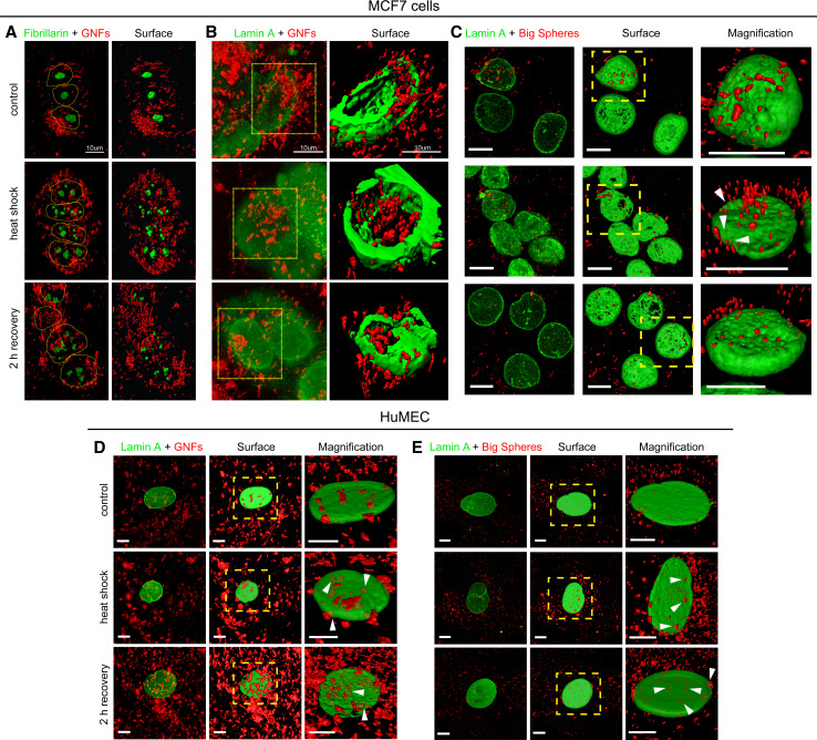
Fig. 6.
a–c Gold nanoflowers disrupt the nucleolar and lamina organization in heat-shocked MCF7 cells. MCF7 cells treated overnight with GNPs were exposed to a 1-h heat shock at 43 °C, followed by 2-h recovery at 37 °C. Cells were fixed, stained with antibodies against fibrillarin or lamin A, and confocal images were acquired as described in the “Materials and methods” section. Gold nanoflowers (GNFs) and big gold nanospheres (big spheres) are pseudocolored in red. Panels show 3D projections of confocal z-stacks. Based on 3D projections of the fluorescent images, surfaces were generated with Imaris software; magnified views are shown as indicated. a MCF7 cells were incubated with gold nanoflowers and fibrillarin was located by indirect immunofluorescence; nuclei are demarcated by dotted lines. b Cells treated with gold nanoflowers or c big gold nanospheres were stained for lamin A. Note that gold nanoflowers associated with nuclei and can be detected in the nuclear interior. Some big gold nanospheres also appear to be located in the nucleus (arrowheads). In cells treated with gold nanoflowers, the distribution of fibrillarin and lamin A indicates severe nucleolar fragmentation and lamina disorganization in response to heat shock; they persist during the recovery period. d, e Association of gold nanoflowers and big gold nanospheres with HuMEC nuclei. The impact of gold nanoflowers and big gold nanospheres on lamin A in HuMEC was analyzed as described for MCF7 cells. The possible presence of GNPs in HuMEC nuclei is marked by arrowheads. All results are representative of at least three independent experiments; size bars are 10 μm

