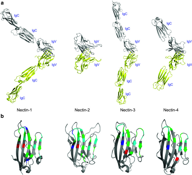
Fig. 5.
Ribbon representations of human nectin homodimers showing the overall structural organizations and interfacial residues at the dimer interfaces. a Individual protomers are colored in yellow and grey. The presented structures of nectin-1 (3ALP) and nectin-3 (4FOM) contain all the three extracellular domains, while the structure of nectin-2 (3R0N) has only the IgV and the structure of nectin-4 (4FRW) has the IgV and only a single IgC. b Ribbon diagram of the IgV domains representing the interfacial residues involved in the homodimerization of each nectin. The residues in red/blue represent charged residues, green represents polar residues, and cyan represents hydrophobic residues

