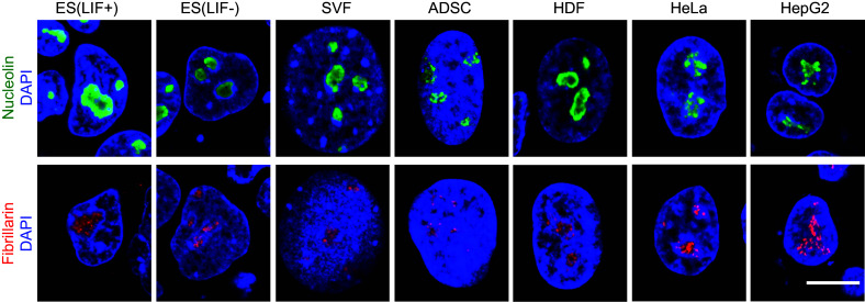
Fig. 1.

Differently sized nucleoli of pluripotent, differentiated, and cancer cells. Mouse ES cells cultured in the presence or absence of LIF, mouse SVF (stromal vascular fraction), human ADSCs (adipose derived stem cells), HDF (human dermal fibroblasts), HeLa cells, and HepG2 cells were stained with antibody to nucleolin (green), fibrillarin (red), and DAPI (blue). Note that pluripotent ES cells (+LIF) and cancer cells (HeLa and HepG2) have large nucleoli compared to differentiated cells (−LIF ES cells, SVF, ADSC, and HDF). Scale bar 10 μm
