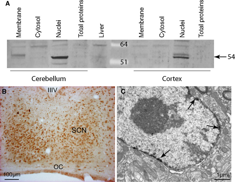
Fig. 1.
Immunodetection of PPARβ/δ in the hamster suprachiasmatic nucleus (SCN). a Specificity for the PPARβ/δ antibody was verified in two hamster cerebral areas (cerebellum and cortex) using Western blotting, which showed the nuclear localization of the protein, and in the liver. b A representative coronal section in the mid-SCN of an adult hamster killed during the light phase. Immunoreactivity was observed throughout the rostro-caudal extent of the SCN with a higher density in the ventro-median part. c An electron microscopy immunolabeling of PPARβ/δ (indicated by the arrows), was only observed in the nuclei of the neurons in the SCN. IIIV third ventricle, OC optic chiasm

