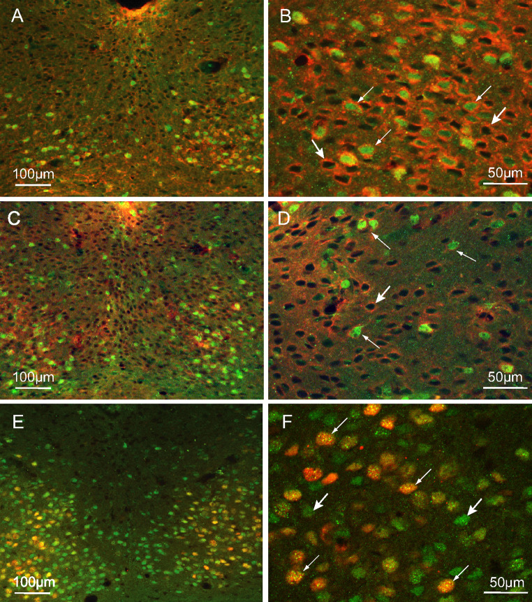
Fig. 5.
Characterization of PPARβ/δ immunoreactive (Ir) neurons. Combined detection of PPARβ/δ (green) and NMDAR1 (red) in the ventral part of the SCN of the hamsters killed at ZT14 (a, b) and in the dorsal part of hamsters killed at ZT06 (c, d); PPARβ/δ is co-expressed with NMDAR1 (thin arrows), there are a number of cells that only expressed NMDAR1 (large arrows). e, f Colocalization of PPARβ/δ (green) and light-induced c-Fos (red) in the SCN of hamsters killed at CT14; the c-Fos-Ir cells are nearly all PPARβ/δ-Ir (thin arrows), and the PPARβ/δ cells are widely distributed within the SCN (large arrows)

