Fig. 1.
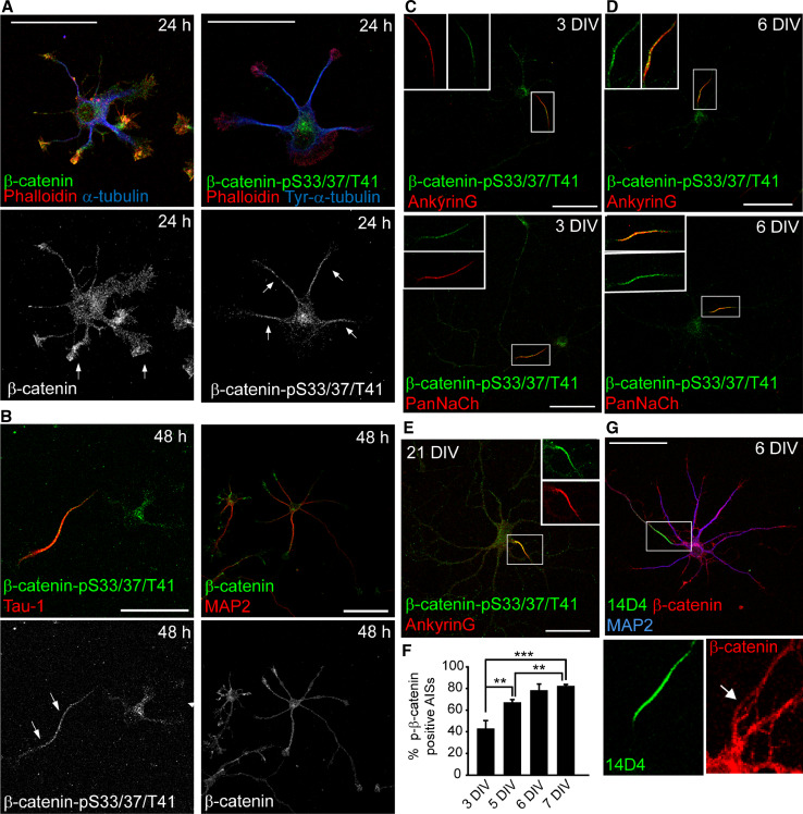
β-catenin-pS33/37/T41 is concentrated at the axon initial segment in cultured hippocampal neurons. a Hippocampal neurons cultured for 24 h were stained for F-actin (red) and antibodies against α-tubulin (blue) and β-catenin (green) in left panels, and tyrosinated-α-tubulin (blue) and pS33/37T41-β-catenin (green) in right panels. Arrows indicate higher β-catenin intensity fluorescence at the growth cone in left panels, while in right panels arrows indicate a tubulin-rich zone with high-intensity fluorescence of pS33/37T41-β-catenin. Note the position of centrosome in neurons labeled with pS33/37T41-β-catenin antibody in the right panel. b Hippocampal neurons cultured for 48 h were stained with pS33/37T41-β-catenin or β-catenin (green) and with tau-1 (axon) or MAP2 (dendrites) antibodies (red). Arrows indicate the axon (c, d) 3-DIV and 6-DIV hippocampal neurons were stained with ankyrinG or PanNaCh (red) and pS33/37T41-β-catenin (green) antibodies. Both proteins are concentrated at the AIS region. Insets show enlarged views of overlapping β-catenin-pS33/37/T41 and AnkyrinG or PanNaCh staining at the AIS. e 21-DIV neurons stained with antibodies against AnkyrinG (red) and β-catenin-pS33/37/T41 (green). Box shows an amplification of the indicated axon initial segment. f Percentage of neurons that show β-catenin-pS33/37/T41 concentrated at the AIS relative to ankyrinG-positive AISs during neuronal differentiation. The graphs represent the mean of three independent experiments (500 neurons/experimental condition in each experiment). **p < 0.01, ***p < 0.001, t test. g Triple immunostaining with β-catenin (red), 14D4 antibody (green), and MAP2 (blue) in 6-DIV neurons. Magnifications show β-catenin staining at the AIS recognized by the 14D4 antibody staining at the AIS. Scale bar 50 µm

